- Khasiat Tanaman Obat, Yohana Arisandi dan Yovita Andriani; ...:Pustaka Buku Murah, 2008. Hlm. 7 - 10
- Trubus Info Kit, Herbal Indonesia Berkhasiat, Bukti Ilmiah dan Cara Racik, Jakarta:Tubus, 2010. Hlm. 154 - 158.
- Herbal penyembuh Impotensi dan Ejakulasi Dini, Joko Suryo; Yogyakarta : B First, 2010. Hlm. 121- 122.
- Obat Tradisional Untuk Pasangan Suami Istri, Hardi Soenanto & Sri Kuncoro S.Sos; Jakarta : PT Elex Media Komputindo, 2009. Hlm. 24 - 26.
- Pengobatan Herbal Ala Nabi, Achmad Rusdi Al-Idrus; Jakarta : Zahra Publishing House, 2009. Hlm.83-84.
- Referensi Online :
Fennel ampuh memperbesar payudara karena memiliki kandungan alami anethole, dianethole dan photoanethole (senyawa yang meningkatkan estrogen dalam tubuh). Fennel juga memiliki phytoestrogen (mirip dengan estrogen) yang menstimulasi pertumbuhan payudara dan meningkatkan produksi ASI bagi ibu menyusui.
Cara Rahasia Menjadi Seperti Perawan Kembali
Selasa, 27 Juli 2010
REFERENSI BIN MUHSIN FENNEL
Minggu, 25 Juli 2010
KHASIAT BIN MUHSIN FENNEL - SYUMIR - ADAS
Minggu, 18 Juli 2010
Epididymis
Epididymis
From Wikipedia, the free encyclopedia
| Epididymis | |
|---|---|
 | |
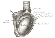
| 1: Epididymis 2: Head of epididymis 3: Lobules of epididymis 4: Body of epididymis 5: Tail of epididymis 6: Duct of epididymis 7: Deferent duct (ductus deferens or vas deferens) | |
 | |
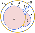
| The right testis, exposed by laying open thetunica vaginalis. | |
| Gray's | subject #258 1242 |
| Vein | Pampiniform plexus |
| Precursor | Wolffian duct |
| MeSH | Epididymis |
The epididymis (pronounced /ɛpɨˈdɪdɨmɪs/, plural: epididymides /ɛpɨˌdɪdɨˈmiːdiːz/) is part of the male reproductive system and is present in all maleamniotes. It is a narrow, tightly-coiled tube connecting the efferent ducts from the rear of each testicle to its vas deferens. A similar, but probably non-homologous, structure is found in cartilaginous fishes.
Contents[hide] |
[edit]Regions
The epididymis can be divided into three main regions
- The head (Caput). The head of the epididymis receives spermatozoa via the efferent ducts of the mediastinum of the testis. It is characterizedhistologically by a thin myoepithelium. The concentration of the sperm here is dilute.
- The body (Corpus)
- The tail (Cauda). This has a thicker myoepithelium than the head region, as it is involved in absorbing fluid to make the sperm more concentrated.
In reptiles, there is an additional canal between the testis and the head of the epididymis, which receives the various efferent ducts. This is, however, absent in all birds and mammals.[1]
[edit]Histology
The epididymis is covered by a pseudostratified epithelium composed of short basal cells and tall principal cells with non-motile stereocilia (long microvilli). The epithelium is separated by a basement membrane from the connective tissue wall which has smooth muscle cells.
[edit]Role in storage of sperm and ejaculant
Spermatozoa formed in the testis enter the caput epididymis, progress to the corpus, and finally reach the cauda region, where they are stored. Sperm entering the caput epididymis are incomplete - they lack the ability to swim forward (motility) and to fertilize an egg. During their transit in the epididymis, sperm undergo maturation processes necessary for them to acquire these functions.[2] Final maturation is completed in the female reproductive tract (capacitation).
During ejaculation, sperm flow from the lower portion of the epididymis (which functions as a storage reservoir). They have not been activated by products from the prostate gland, and they are unable to swim, but are transported via the peristaltic action of muscle layers within the vas deferens, and are mixed with the diluting fluids of the seminal vesicles and other accessory glands prior to ejaculation (forming semen).
The epididymis possesses numerous, long atypical microvilli. These processes are often called stereocillia; this is incorrect, as they neither contain the microtubular structures of cilia nor function like cilia.[3]
[edit]Pathology
An inflammation of the epididymis is called epididymitis.
[edit]Embryology and vestigial structures
A Gartner's duct is a homologous remnant in the female.
In the embryo, the epididymis develops from tissue that once formed the mesonephros, a primitive kidney found in many aquatic vertebrates. Persistence of the cranial end of the mesonephric duct will leave behind a remnant called the appendix of the epididymis. In addition, some mesonephric tubules can persist as the paradidymis, a small body caudal to the efferent ductules.
[edit]Epididymectomy
This is the surgical removal of the Epididymis carried out under local anaesthesia. This is most often performed to relieve pain associated post-Vasectomy.
[edit]Gallery
Micrograph of an epididymis. H&E stain. | |||
[edit]Notes
- ^ Romer, Alfred Sherwood; Parsons, Thomas S. (1977). The Vertebrate Body. Philadelphia, PA: Holt-Saunders International. pp. 394–395. ISBN 0-03-910284-X.
- ^ Jones R (1999). "To store or mature spermatozoa? The primary role of the epididymis". Int J Androl 22 (2): 57–67. doi:10.1046/j.1365-2605.1999.00151.x. PMID 10194636. abstract
- ^ Stevens, Alan; Lowe, James N. (2005). Human histology. Philadelphia: Elsevier Mosby. ISBN 978-0-323-03663-4.
[edit]External links
- Histology at BU 16903loa
- inguinalregion at The Anatomy Lesson by Wesley Norman (Georgetown University) (testes)
| ||||||||||||||||||||||||||||||||||


